
Hand Tumours
Stop Hand Tumours Pain
Tumours of hand bones and fingers can be of wide description such as lumps, nodules. They are usually not cancerous, but they are usually unsightly.

Tumours of hand bones and fingers can be of wide description such as lumps, nodules. They are usually not cancerous, but they are usually unsightly. Patient usually comes with complaint of swelling with unaesthetic appearance or could have sensory or motor symptoms due to compression of the underlying nerves. Due to complex movements of hand and fingers, treatment of tumours of hand is very important more so in some professions like musician, typist, military services.
Tumours of the hand can arise from the skin, fat, tendons (structures which moves the fingers), bone, muscles, nerves and the blood vessels.
Of all the hand tumours, few tumours are very common-
- Cysts
- Lipoma
- Ganglion
- Glomus Tumor
- Giant cell tumour of the tendon sheath
- Neurofibroma
- Sarcomas or soft tissue tumours
- Bone tumour
Cysts
Cysts are very common tumours of the hand.
The most common cysts found in the hand are sebaceous cyst or inclusion cyst.
Both the cysts are harmless, do not create any problem apart from difficulty in doing daily tasks because of their bulky nature and aesthetic concern.
They should be excised completely otherwise recurrences are common.
Lipoma
Lipoma is the tumor of fat cells located just below the skin of the hand. They are often confused with the Cysts. They are non-painful but can be if the underlying nerve is compressed. They also do not create any problem apart from difficulty in doing daily work because of their bulky nature and aesthetic concern.
Lipoma on the hand should be excised but if on forearm and arm, can be left.
If multiple lipomas are present on arm and forearm region, they can be left as such and only big ones which cause cosmetic concerns are removed.
Ganglion
Ganglion is the most common soft tissue tumor of the hands. They are filled with mucus and are cystic in nature and often develops from capsule of the joint or tendon sheath.
Ganglions are most commonly found on the back of the hand at the wrist joint. They can also be found at the front of the hand at the wrist joint.
Ganglions usually grow slowly and do not cause any pain until large enough but they interfere with the movements of the wrist. They can be quite painful at extreme movements of the wrist joint while working.
Ganglions are readily diagnosed by clinical examination. They are present as a small bumpy swelling on the back or front of wrist.
The diagnosis and deeper communication or origin (whether joint or tendon sheath) can be confirmed by ultrasonography or MRI Even mildly painful Ganglion cysts should be removed when they are diagnosed to prevent the complication of spontaneous rupture which can lead to spread of mucinous material along the tendon sheath in the forearm.
Complete excision by surgery is the best form of treatment for ganglion which should be done under Brachial block or General Anaesthesia.
No form of aspiration of the cyst or puncture of the cyst with the needle or steroid injection should be given in the cyst as it can lead to further complications.
Recurrence is common.
Glomus Tumor
Glomus tumours are very commonly found in women in their middle age years.
These tumours are non-cancerous, very small and vascular (made up of blood vessels and highly sensitive nerve endings)
They are found beneath the nail plate and nail bed of the fingers.
They are very painful and often go undiagnosed by most of the clinicians. Patient always complaints of severe pain in the affected finger and hand. Glomus tumor often presents with a classical triad of symptoms - severe pain, point tenderness and cold sensitivity. Few can have bluish discoloration of the nail or deformity of the nail along with the above symptoms.
X rays and imaging studies are mostly normal. Glomus tumours are most commonly diagnosed with MRI scan of the affected finger.
Surgical excision is the treatment of choice and usually give relief to most of the patients.
The surgery is usually done under brachial block anesthesia or general anaesthesia. The nail plate is removed, nail bed is slit longitudinally and the tumor is removed. The nail bed is sutured with absorbable sutures.
Giant cell tumor of the Tendon sheath
Giant cell tumour of the tendon sheath are also very common tumours of the hand. They are also non-cancerous and are present along the joints, ligaments and tendon sheaths.
They are firm, nodular and non-painful.
They can grow to much larger dimension, and these usually present with symptoms of nerve compression or unaesthetic appearance.
The diagnosis is usually confirmed with MRI scan and Fine Needle Aspiration Cytology (FNAC).
Complete excision of this tumor is must while preserving the nerve and artery of the fingers.
Schwannoma and Neurofibroma of the Upper Limb
Schwannoma is the tumor of the outer covering of the nerves.
Neurofibroma is the tumor of nerve fibres within the nerve.
Schwannoma is the most common benign nerve tumour of the upper limb. They are well circumscribed, well defined, slow growing mass developed along the course of one of the major nerves of the arm (median, ulnar or radial). Very rarely, you get neurological deficit of the involved nerve.
On clinical examination, if the mass is pressed it can cause radiation pain in the area of distribution of the nerve. The diagnosis is usually confirmed with MRI scan and Fine Needle Aspiration Cytology (FNAC).
Surgical treatment is the treatment of choice and excision is done with preservation of the nerve.
Neurofibroma is a slow growing tumor of the nerve fascicles within the substance of the nerve. They can develop in any of the nerves of the arm and can present with neurological deficit also. They can be painful too if compressed.
The diagnosis is usually confirmed with MRI scan and Fine Needle Aspiration Cytology (FNAC).
In few cases, multiple neurofibroma are seen in contrast to schwannoma which is mostly single.
Surgical treatment is the treatment of choice and excision is done with nerve reconstruction and nerve grafting.
Soft tissue Sarcomas of hand and Upperlimb
Soft tissue sarcomas are better known as cancers of the soft tissue. The most common soft tissue sarcomas of the hand are malignant fibrous histiocytoma, epithelioid sarcoma and synovial sarcoma which are usually diagnosed on biopsy or FNAC.
They usually present as rapidly growing growth or mass over the hand or fingers with areas of hardness or ulceration, mass in an atypical location with enlarged lymph nodes in the axilla.
Investigations
- Radiograph of hand to see bone erosion.
- HR Ultrasonography of swelling in few cases
- NCV/ EMG (if there is any nerve compression)
- MRI / CT scan of hand
- Chest Xray, CT scan of the chest and axilla to see whether it has spread to lungs and lymph nodes or not.
Treatment
Soft tissue sarcomas are usually treated with wide local excision of the swelling with biopsy of swelling. The defect created by the excision of the sarcoma is reconstructed with an appropriate reconstructive technique as discussed in limb salvage surgery.
Biopsy determine the next step in management.
It tells whether the tumour is benign or malignant or which subtype of soft tissue sarcoma is present.
If benign then no other intervention needed but monitoring and follow up is needed to assess the recurrence of tumour.
If malignant, secondary procedure or adjunct therapy in form of chemotherapy or radiotherapy may be required.
Bone tumors of hand
Bone tumors of hand are not very common and could be benign or cancerous. The most common bone tumors of the hand are enchondroma, chondroma, osteochondroma, exostoses, osteoid osteoma, giant cell tumours of the bone, aneurysmal bone cyst, Ewing's sarcoma and osteosarcoma. They are usually diagnosed on biopsy or FNAC.
They usually present as rapidly growing growth or mass over the hand or fingers with areas of hardness or ulceration, with or without enlarged lymph nodes in the axilla.
Investigations
- Radiograph of hand to see whether the mass is rising from the bone or not
- HR Ultrasonography of swelling to be done in few cases
- MRI / CT scan of hand easy gold standard investigating technique to look for the extent off the tumor and its soft tissue extension
- FNAC is done to know its subtype.
- Chest Xray, CT scan of the chest and axilla to see whether it has spread to lungs and lymph nodes or not.
Treatment
Bone tumours of the hand are usually treated with wide local excision of the tumor with preservation of vital important structures. The defect created by the excision of the tumor is reconstructed with an appropriate reconstructive technique as discussed in limb salvage surgery.
In few cases where reconstruction is not possible or fruitful in malignant or cancerous bone tumors, then amputation of the finger or hand is advised to save the life of the patient and to limit the possible spread of the tumor to various parts of the body.
Biopsy determine the next step in management.
It tells whether the tumour is benign or malignant or which subtype of bone tumor.
If benign then no other intervention needed but monitoring and follow up is needed to assess the recurrence of tumour.
If malignant, secondary procedure or adjunct therapy in form of chemotherapy or radiotherapy may be required.
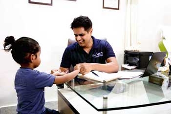
Hand Tumours Images
Indivisual results may vary from person to person.
These pictures are shown for the purpose of education only.


Shaping dreams through


Know your surgeon better
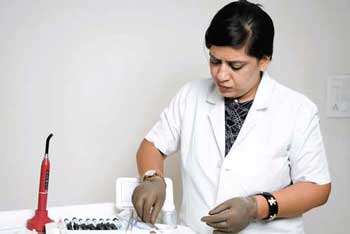
Best plastic surgeon, Dr. Amit Agarwal is an American Board Certified, extensively trained, and best Plastic & Aesthetic surgeon in Lucknow. He is the Chief Plastic Surgeon heading the Department of Plastic, Microvascular, and Craniofacial surgery at Vivekananda Polyclinic and Institute of Medical Sciences, Lucknow, U.P, India. He maintains a busy practice at Avadh and Nishat Hospital and his own center - Kayakriti Plastic Surgery & Dental Center. He was formerly a Consultant in the Department of Plastic Surgery and Burns at the prestigious SGPGI, Lucknow.
MS, DNB (General Surgery) MCh, DNB (Plastic Surgery),
MNAMS, FACS, FICS, FRCS (Edinburgh, UK)
His Credentials


Three pillars of kayakriti
Privacy
We believe your experience with us should be comfortable and hassle-free to make it one of your best lifetime experiences for yours. We, here at the clinic, take full precautions to maintain your privacy in any manner. We also provide a staff who will receive you from the gate and take you to the chamber directly if you demand.
Trust
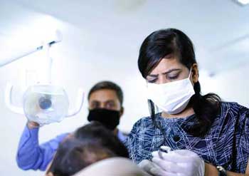
Our Surgeon is highly qualified and internationally certified with a team of skilled staff to perform any surgical or non-surgical treatment on your body.
Safety
When you plan to undergo any surgery you should always keep in mind that it's your body and it's a surgery. We, here always keep your safety a priority and will never recommend you to undergo any such procedure which is not safe for you. We also provide you with a detailed description of the complications which may occur after the surgery during the consultation as it's a surgical procedure so there may be some complications depending on the way your body reacts.
Kayakriti in news


Frequently Asked Questions
If you have flat or small breast and you want to improve your breast and hip contour ratio then you are a good candidate for it. The answer will be best provided after the first consultation with Dr Amit Agarwal.
Acute pain will be there for almost a week which gradually reduces and there will be soreness and swelling which may take up to 3 weeks to subside.
You can join your work and daily routines after a week of the procedure and can start exercising after 3 weeks of it.
Yes, you have to wear it round the clock unless we suggest you to remove it.
This surgery does not affect the ducts or the areas of the breast involved in milk production. Thus, it does not affect the breast feeding.
This surgery does not affect the ducts or the areas of the breast involved in milk production. Thus, it does not affect the breast feeding.
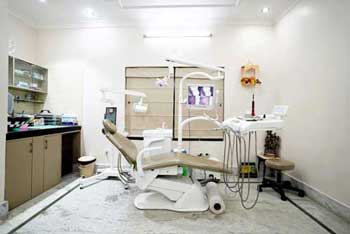
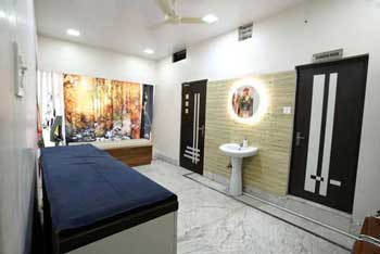


Kayakriti Plastic Surgery & Dental Center
D-43, Near Punjab National Bank, Rajajipuram, Lucknow, Uttar Pradesh - 226017, India
Phone No. +919695940009, +919695940006
Map Location

Social Media Presence